Biography
I obtained my PhD in 2010 from Rennes University (with F. Tiaho), where I studied xenopus larvae with 2-photon microscopy. I then joined the group of N. Peyriéras (Gif-sur-Yvette) to investigate cell dynamics in live zebrafish embryos, sharpening my skills to babysit embryos to make sure they are happy and comfortable under the scope. I joined the group of M. Zernicka-Goetz (Cambridge, UK), where I learnt techniques related to mammal embryos. There I realized how demanding these bunch of cells are when it deals with microscopy. I met 3D in vitro systems, and moved to Bordeaux in 2015 to work with P. Nassoy and the alginate capsules! Here, as a CNRS researcher, I study these balls of cells, and still develop microscopy approaches. It was obvious to me to muster up the techniques specifically developed by each community and that's how the "Mount(n)" was born.Research topics
Whether on embryos or on in vitro systems, I study cell self-organisation in a 3D environment using biophysics approaches.Not only looking at how cells dynamically organise to give rise to a bigger structure, but also using that knowledge to make them organize in a specific manner, trying to achieve what we call a physiomimetic morphogenesis.
Fields of interest
Cell self-organization; tissue biomechanics; physiomimetic morphogenesis
Technologies
Applications
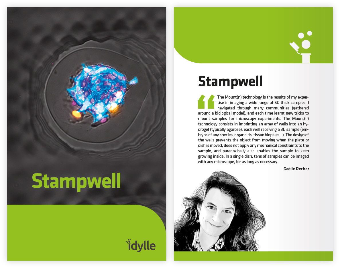
Stampwell
The first stamp to imprint arrays of wells in hydrogels and to mount 3D biological samples for parallelized microscopy experiments. Stampwell ensures the sample stillness, long-term medium-throughput and automatized multi-position image acquisition for a significant timesaving.
Several publications
Andrique, L*, Recher, *, Alessandri, K, Pujol, N, Feyeux, M, Bon, P, Cognet, L, Nassoy, P, and Bikfalvi, A (2019). A model of guided cell self-organization for rapid and spontaneous formation of functional vessels. Science Advances, eaau6562
https://doi.org/10.1126/sciadv.aau6562
Shahbazi M, Scialdone A, Skorupska N, Weberling A, Recher G, Zhu M, Jedrusik A, Devito L, Noli L, Macaulay I, Buecker C, Khalaf Y, Ilic D, Voet T, Marioni J, Zernicka-Goetz M (2017) Pluripotent state transitions coordinate morphogenesis in mouse and human embryos. Nature
https://doi.org/10.1038/nature24675
Alessandri K*, Andrique L*, Feyeux M*, Bikfalvi A, Nassoy P & Recher G† (2017) All-in-one 3D printed microscopy chamber for multidimensional imaging, the UniverSlide. Scientific Reports
https://doi.org/10.1038/srep42378
Joly J-S, Recher G, Brombin A, Ngo K & Hartenstein V (2016) Conveyor-belt neurogenesis: a conserved mode of generating a complex, homotopically patterned visual system in insects and vertebrates. Current Biology
https://doi.org/10.1016/j.cub.2016.08.017
Shahbazi M*, Jedrusik A*, Vuoristo S*, Recher G, Hupalowska A, Campbell A, Fishel S & Zernicka-Goetz M (2016) Self-organization of the human embryo in the absence of maternal tissues. Nature Cell Biology
https://doi.org/10.1038/ncb3347
Faure E*, Savy T*, Rizzi B*, Melani C*, Stasova O*, Fabrèges D*, Spir R, Hammons M, Cunderlik R, Recher G, Lombardot B, Duloquin L, Colin I, .../..., Peyriéras N & Bourgine P (2016) An algorithmic workflow for the automated processing of 3D+time microscopy images of developing organisms and the reconstruction of their cell lineage. Nature Communications
https://doi.org/10.1038/ncomms9674
Recher G*, Jouralet J*, Brombin A, Heuzé A, Mugniery E, Hermel J-M, Desnoulez S, Savy T, Herbomel P, Bourrat F, Peyriéras N, Jamen F & Joly J-S (2013) Zebrafish midbrain slow-amplifying progenitors exhibit high levels of transcripts for nucleotide and ribosome biogenesis. Development
https://doi.org/10.1242/dev.099010
Rouède D, Bellanger J-J, Schaub E, Recher G & Tiaho F (2013) Theoretical and Experimental SHG Angular Intensity Patterns from Healthy and Proteolysed Muscles. Biophysical Journal
https://doi.org/10.1016/j.bpj.2013.02.047
Recher G, Rouède D, Schaub E & Tiaho F (2011) Skeletal muscle sarcomeric SHG patterns photo-conversion by femtosecond infrared laser. Biomedical Optics Express
https://doi.org/10.1364/BOE.2.000374